Have you received a breast density notification in your mammogram results letter? Being told that you have dense breast tissue can be confusing and even a little scary. While having dense breast tissue is common and not considered an abnormality, it can pose additional risk when it comes to breast cancer. For this reason, the FDA recently issued a national requirement stating that all breast imaging facilities must notify women if they have higher levels of dense tissue (50% or greater). Women’s Imaging Specialists believes that prevention begins with knowledge. We have been committed to educating patients about dense breast tissue and the additional imaging options available for nearly a decade, long before it became a national law.
What is Dense Breast Tissue?
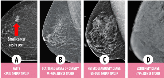
Having dense breast tissue means that your breasts have less fatty tissue and more glandular and connective tissue. Dense breast tissue cannot be determined by how a breast looks or feels, it is only determined after mammogram images are reviewed. On a mammogram, fatty breast tissue appears dark and transparent, making it easier to detect cancer and other potential abnormalities. Dense breast tissue blocks x-ray penetration and therefore shows up white on a mammogram, the same color that cancer appears. Because dense tissue and cancer both show up white on a mammogram, the ability to detect breast cancer in highly dense women more difficult. The higher level of density your breasts have, the more difficult it becomes. Breast density is classified into four levels.
Level A – Mostly fatty with less than 25% of the breast tissue being dense
Level B – Fatty breast tissue with 25 – 50% scattered density
Level C – Heterogeneously dense, 50 – 75% of the breast tissue is dense
Level D – Extremely dense, more than 75% of the breast tissue is dense
Breast density can change throughout a woman’s life due to various factors. The most common factor is age. Woman typically have higher level of density in their breasts when they are younger. For some women, that density declines around menopause. Other factors that can affect breast tissue include hormone therapy, body weight, pregnancy, and genetics.
Does Having Dense Breast Tissue Increase My Risk of Breast Cancer?
If you are told that you have dense breast tissue after your mammogram, it does not mean that your mammogram is abnormal. Dense breast tissue is common. In fact, over 40% of women between the ages 40 – 74 have level C or D dense breast tissue. However, having heterogeneously or extremely dense tissue does put you at a higher risk for developing breast cancer. Women with dense tissue are 4 to 6 times more likely to develop breast cancer than those with fatty breast tissue. In addition, the masking effect caused by the density reduces a mammogram’s sensitivity, making it much more challenging to diagnose breast cancer at an early stage.
Are There Additional Adjunct Imaging Options Available to Women with Dense Breast Tissue?
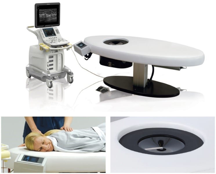
Women’s Imaging Specialists is proud to be one of the only breast imaging facilities in the Southeast to offer the Automated 3D Whole Breast Ultrasound to patients with category C or D density. This ultrasound is the only adjunct test that is specifically designed for women with dense breast tissue. It utilizes sound waves to penetrate through the dense tissue and scans the entire breast. Patients are able to lie in a comfortable prone position with no compression, or radiation used, and the scan takes just 30 seconds per breast. The 3D Whole Breast Ultrasound provides 3D volumetric images of the breast that can allow radiologists to see things that may have been missed on a mammogram and is a much more cost-effective alternative to an MRI for women with no other high-risk factors.
Can I Have an Automated 3D Whole Breast Ultrasound Instead of a Mammogram?
Regardless of your density, screening mammography is still the best tool to detect breast cancer at an early and treatable stage. A 3D Whole Breast Ultrasound is not a replacement for an annual mammogram, it should only be considered as supplemental screening.
Should I Have a 3D Whole Breast Ultrasound if I Have a Family History or Other High-Risk Factors?
The 3D Whole Breast Ultrasound is a much more cost-effective alternative to an MRI for woman who have no other high-risk factors or family history. If you are high-risk or have a strong family history of breast cancer, your healthcare provider may recommend an MRI or additional screenings.
Bottom Line
Having a better understanding of dense breast tissue and the risks it can pose, can not only help you actively participate in discussions with your healthcare provider, and make informed decision about the imaging options best for you. It can also help to alleviate anxiety and ensure that you are receiving the best possible care! If you have questions or would like to learn more about the Automated 3D Whole Breast Ultrasound, please explore our services page, or call a Women’s Imaging Specialists location near you!
