Screening mammography has been evolving over the last 100 years, resulting in a 20-30% reduction in breast cancer -associated mortality rate. Even today, there continues to be advances in technology, making screenings more accurate and less stressful. The most recent advancement, approved in 2011, was the introduction of 3D mammography, also known as digital breast tomosynthesis (DBT).
When interpreted by trained radiologists, this new technology is credited in assisting in the earlier and more accurate detection of possible breast cancers, while also reducing the number of women who are called back for additional testing.
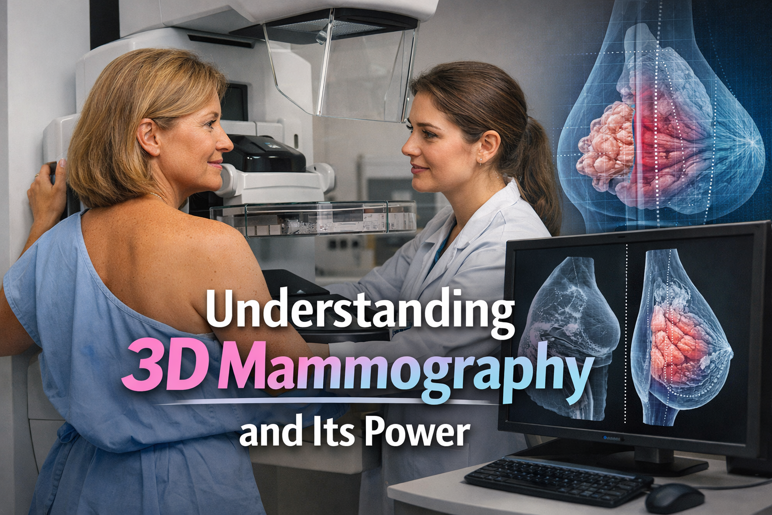
What is 3D mammography?
Traditional 2D mammography takes one flat image of the breast from the front and the side. While effective, these images can sometimes be hard to interpret because breast tissue overlaps, which can hide small cancers or create areas that look suspicious but are actually normal.
3D mammography works differently. During the exam, the machine takes multiple low-dose X-ray images from different angles. A computer then combines these images to create a series of very thin layers of the breast. The radiologist can scroll through these layers one at a time, similar to flipping through pages in a book. This layered view makes it easier to see what is really there.
What is the experience like for patients?
For most patients, a 3D mammogram feels very similar to a standard mammogram.
- The breast is positioned and gently compressed for a few seconds.
- The machine moves slightly to capture multiple images.
- The exam usually takes only a few minutes longer than a 2D mammogram.
Both the 2D and the 3D images play an important role in the interpretation of the mammogram. As part of our services, Women’s Imaging Specialists will create a synthetic 2D image from the 3D data. This allows doctors to review both views without significantly increasing radiation exposure to the patient.
How is 3D mammography used?
3D mammography is used in two main ways:
Screening mammograms
These are routine exams for people who do not have symptoms. Large studies show that using 3D mammography for screening finds more cancers and reduces unnecessary call-backs.
Diagnostic mammograms
If a patient has a symptom or an abnormal screening result, 3D mammography can be used to take a closer look. It is often combined with ultrasound or MRI when needed.
How 3D Mammography Improves Breast Cancer Detection
It reduces tissue overlap
In 2D images, normal breast tissue can overlap and hide small tumors. By looking at the breast layer by layer, 3D mammography reduces this problem and helps doctors spot cancers that might otherwise be missed.
Reducing the need for additional imaging
In traditional 2D mammography, overlapping breast tissue can obscure small tumors, making them harder to detect. This challenge is especially common in women with dense breast tissue, where both normal tissue and cancer appear white on a mammogram. By capturing the breast tissue in thin, layered images, 3D mammography reduces tissue overlap and provides a clearer view. This helps radiologists identify cancers that might otherwise be missed. For patients with extremely dense breast tissue, a 3D whole breast ultrasound used in conjunction with a 3D mammogram may provide additional benefit in detecting breast cancer.
What does the research show?
Higher cancer detection rates
Studies consistently show that 3D mammography detects up to 40% more breast cancers than 2D mammography. i. McDonald ES et al. Effectiveness of Digital Breast Tomosynthesis Compared With Digital Mammography. JAMA Oncology, 2016.
- A major review of multiple studies found that 3D mammography detected about 9 cancers per 1,000 women screened, compared to about 6 per 1,000 with 2D mammography.
Earlier detection of invasive cancers
Studies published in major medical journals such as omosynthesis vs Digital Mammography Screening in Women with a Family History of Breast Cancer,” published in JAMA Oncology have found that when breast cancers are detected with 3D mammography, they are more likely to be found at an early, localized stage, making them more treatable and significantly improving the likelihood of a positive outcome.
Finding cancer at an earlier stage often leads to more treatment options and better outcomes.
Who benefits most from 3D mammography?
While 3D mammography provides clearer imaging and benefits everyone, it may be especially helpful for
- Women with dense breast tissue
- Women in their 40s and early 50s
- Patients who have been called back in the past due to unclear 2D images
The bottom line
3D mammography is a major advancement in breast cancer screening. It helps doctors:
- Detect more cancers during routine screenings
- Identify cancers earlier, when they are often easier to treat
- Reduce unnecessary call-backs and follow-up testing
At Women’s Imaging Specialists, 3D mammography is offered as the standard for all mammograms, providing the clearest, most detailed imaging for every patient. You can schedule your next mammogram at one of our 17 convenient locations by visiting our website or calling 866-300-8512.
References
- Friedewald SM et al. Breast Cancer Screening Using Tomosynthesis in Combination With Digital Mammography. JAMA, 2014.
- Ciatto S et al. Integration of 3D Digital Mammography With Tomosynthesis for Population Breast-Cancer Screening. Radiology, 2013.
- Skaane P et al. Comparison of Digital Mammography Alone and Digital Mammography Plus Tomosynthesis in a Population-based Screening Program. Radiology, 2013.
- U.S. Food and Drug Administration (FDA). Mammography Quality Standards Act and Digital Breast Tomosynthesis.
- McDonald ES et al. Effectiveness of Digital Breast Tomosynthesis Compared With Digital Mammography. JAMA Oncology, 2016.
