


Many people think breast cancer will always cause a noticeable lump, pain, or visible change before it becomes a problem. In reality, breast cancer can develop quietly and may not show any symptoms for months or even years. This is one reason routine screening is so important, as it supports early breast cancer detection before symptoms develop.
Modern breast imaging technologies help doctors find changes in breast tissue long before they can be felt or seen. Imaging can spot small abnormalities, subtle tissue changes, and early signs of disease that may not cause any symptoms.
Detecting breast cancer early creates more chances for timely treatment and may lessen the need for more extensive procedures later.
Whether you are scheduling your first screening mammogram or exploring available imaging options, understanding how these tools work is important. It can help you make informed choices about your breast health.
Why Breast Cancer Often Develops Before Symptoms Appear
Did you know breast cancer begins at the cellular level? During its earliest stages, abnormal cells may be too small to create symptoms or physical changes.
With this information in mind, it’s important to remember that feeling healthy does not necessarily mean breast tissue is free of abnormalities. Screening often identifies many breast cancers before a lump becomes noticeable.
According to the American Cancer Society, screening is designed to detect cancer before symptoms appear, when treatment may be more effective and less invasive. Routine imaging supports early breast cancer detection by helping providers evaluate internal breast structures that cannot be assessed through self-awareness or physical examination alone.
This is where mammograms come in, providing reassurance through advanced imaging solutions.
The Role of Mammograms in Early Breast Cancer Detection
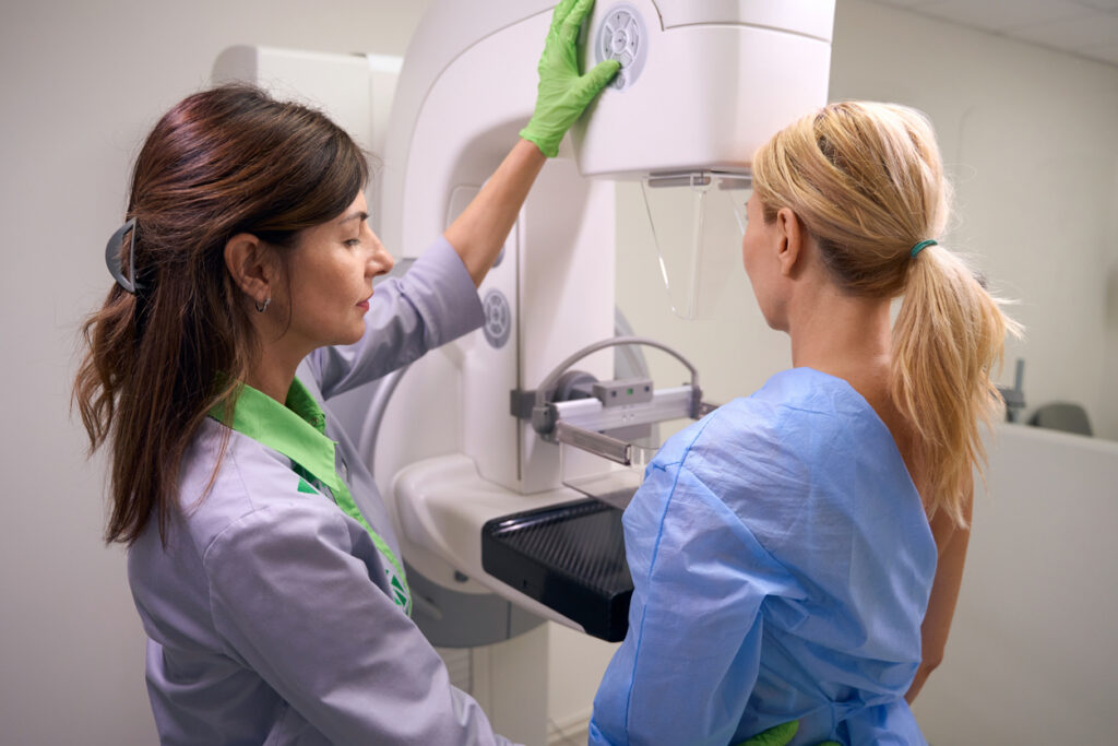
Mammography remains one of the most widely used and effective breast screening tools available today. For early detection of abnormalities, this tried-and-true method is the most accurate.
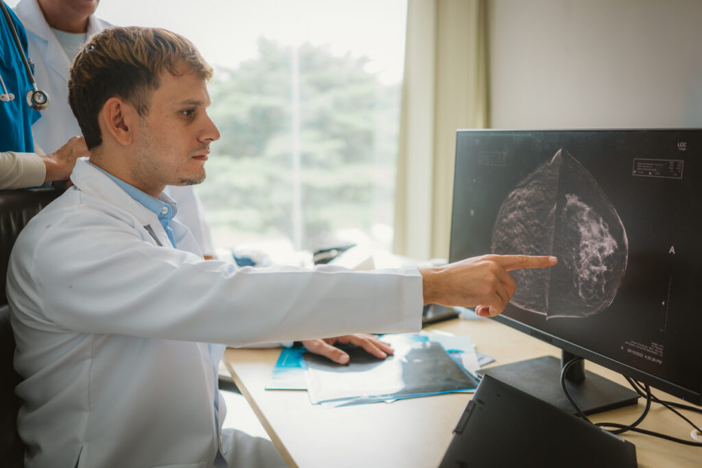
A mammogram uses low-dose X-ray imaging to detect suspicious findings such as
- Small masses
- Tiny calcium deposits (microcalcifications)
- Tissue distortions
- Areas of asymmetry
Many of these findings are impossible to detect through touch alone, even if you’re great about keeping up with physicals. Screening mammograms are specifically designed for people without symptoms and play a central role in early breast cancer detection.

The American Cancer Society recommends discussing screening schedules with your provider based on age, personal history, and risk factors.
What Breast Imaging Can Detect Before You Notice Changes
Breast imaging can identify subtle abnormalities that develop long before symptoms appear, such as
- Small tumors not yet large enough to feel
- Clusters of abnormal calcifications
- Architectural distortion within tissue
- Early-stage lesions
- Areas requiring closer monitoring
These findings do not automatically mean cancer is present, but they may indicate the need for additional imaging or evaluation. That ability to identify concerns before symptoms appear is one of the greatest benefits of screening.
How 3D Mammography Improves Cancer Detection
3D mammography, also called digital breast tomosynthesis, creates multiple images of the breast from different angles. Instead of viewing tissue as a single flat image, radiologists can evaluate thin image layers for improved visibility.
Potential benefits of 3D mammography include:
- Better visualization of overlapping tissue.
- Improved detection in certain patients.
- Fewer call-backs for additional imaging.
- More detailed evaluation of suspicious areas.
For many patients, especially those with dense breast tissue, 3D imaging may provide additional clarity during screening.
When Breast Ultrasound May Be Recommended
Breast ultrasound uses sound waves rather than radiation to evaluate breast tissue.
Ultrasound is commonly recommended:
- After an abnormal mammogram
- To evaluate a palpable concern
- For patients with dense breast tissue
- To help distinguish fluid-filled versus solid findings
Breast ultrasound does not replace mammography but may serve as an important supplemental tool depending on clinical findings and breast density.
How Breast MRI Supports High-Risk Screening
Breast MRI creates highly detailed images using magnetic fields and contrast imaging. Providers may recommend a breast MRI for:
- Patients with an elevated lifetime breast cancer risk
- Strong family history
- Certain genetic factors
- Supplemental screening alongside mammography
MRI is particularly useful because it can identify abnormalities that may not appear clearly on mammography alone.
The American Cancer Society notes that MRI may be recommended alongside mammograms for select higher-risk patients.
The Role of ABUS for Women With Dense Breast Tissue
Automated Breast Ultrasound (ABUS) is designed to improve imaging evaluation for women with dense breast tissue.
Dense tissue can make abnormalities harder to visualize using standard mammography alone.
ABUS offers:
- Supplemental whole-breast imaging
- Standardized image acquisition
- Additional visibility in dense tissue
- Improved confidence during screening evaluation
For some patients, ABUS may become an important part of a personalized screening strategy.
Why Early Detection Leads to Better Outcomes
Finding breast cancer earlier often provides more treatment options and may reduce treatment complexity.
When doctors detect cancer at an earlier stage, patients may have access to more treatment options, including the following:
- Smaller surgical procedures
- Earlier intervention
- Targeted treatment approaches
- Reduced treatment intensity
Early detection does not guarantee outcomes, but screening helps identify concerns sooner than waiting for symptoms to develop.
The American Cancer Society emphasizes that regular screening remains one of the most reliable ways to find breast cancer early.
When to Schedule Routine Breast Cancer Screening
Breast cancer screening recommendations vary depending on age, personal risk factors, family history, and overall health. Many average-risk individuals begin discussing routine mammograms around age 40, though individual recommendations may differ.
Of course, getting your primary care physician’s take on when to begin screening is the best timeline to operate by.
If you are unsure when to begin screening, talk with your healthcare provider about:
- Family history
- Breast density
- Prior imaging results
- Personal risk factors
Staying consistent with recommended screening intervals is often more important than waiting for symptoms to appear.
Taking a Proactive Approach to Breast Health
Breast health is not only about responding to symptoms; it is also about staying proactive. Patients can make educated healthcare decisions, find peace of mind, and detect changes earlier with routine breast imaging.
Mammography, breast ultrasound, MRI, and ABUS each play an important role in supporting early breast cancer detection. Even when you feel healthy and notice no changes, screening remains one of the most valuable tools available for protecting long-term breast health.
Stay current with your preventive care plan and reach out to our team to schedule your next imaging appointment.
First mammogram? Get the scoop on how to prepare for your mammogram appointment.